108605
论文已发表
注册即可获取德孚的最新动态
IF 收录期刊
- 3.4 Breast Cancer (Dove Med Press)
- 3.2 Clin Epidemiol
- 2.6 Cancer Manag Res
- 2.9 Infect Drug Resist
- 3.7 Clin Interv Aging
- 5.1 Drug Des Dev Ther
- 3.1 Int J Chronic Obstr
- 6.6 Int J Nanomed
- 2.6 Int J Women's Health
- 2.9 Neuropsych Dis Treat
- 2.8 OncoTargets Ther
- 2.0 Patient Prefer Adher
- 2.2 Ther Clin Risk Manag
- 2.5 J Pain Res
- 3.0 Diabet Metab Synd Ob
- 3.2 Psychol Res Behav Ma
- 3.4 Nat Sci Sleep
- 1.8 Pharmgenomics Pers Med
- 2.0 Risk Manag Healthc Policy
- 4.1 J Inflamm Res
- 2.0 Int J Gen Med
- 3.4 J Hepatocell Carcinoma
- 3.0 J Asthma Allergy
- 2.2 Clin Cosmet Investig Dermatol
- 2.4 J Multidiscip Healthc

C14orf159 通过灭活 ERK 信号传导抑制胃癌细胞的侵袭和增殖
Authors Zhu YM, Li Q, Gao XZ, Meng X, Sun LL, Shi Y, Lu ET, Zhang Y
Received 10 June 2018
Accepted for publication 17 January 2019
Published 19 February 2019 Volume 2019:11 Pages 1717—1723
DOI https://doi.org/10.2147/CMAR.S176771
Checked for plagiarism Yes
Review by Single-blind
Peer reviewers approved by Dr Justinn Cochran
Peer reviewer comments 2
Editor who approved publication: Dr Kenan Onel
Background: C14orf159,
a new protein, has been identified recently. But its expression in tissues and
clinicopathologic correlation is still unknown.
Patients and methods: We
carried out immunohistochemistry staining in 144 gastric cancer cases in this
study. Then Western blot was used to detect the expression of protein. MTT and
matrigel invasion assay were used to assess the biological effects.
Results: The
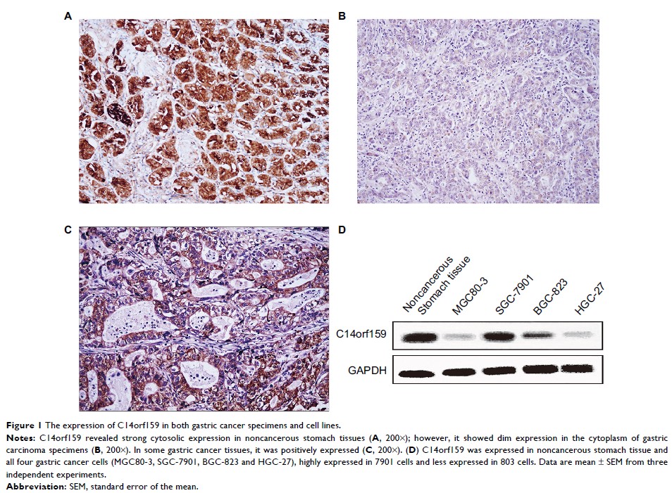
immunohistochemical results indicated that the expression of C14orf159 in
normal gastric mucosa close to cancer tissue was remarkably higher than that in
stomach carcinoma samples (63.9% and 34.7%, respectively, P <0.001).
Negative C14orf159 expression was dramatically related to high TNM stages (P =0.033) and
positive lymph node metastasis (P =0.008). Once C14orf159 was overexpressed, the
expression levels of phosphorylated ERK and its regulated downstream molecules,
such as Snail, phosphorylated P90RSK and Cyclin D1, were decreased, while the
expression level of E-cadherin was increased. Finally, the invasion and
proliferation capacity of gastric cancer cells was inhibited.
Conclusion: In other
words, loss of C14orf159 is associated with the progression of gastric cancer.
The role of C14orf159 in repression of proliferation and invasion may be due to
resuming E-cadherin and abolishing Snail and Cyclin D1 expression through
inactivating ERK–P90RSK pathway.
Keywords: C14orf159,
gastric cancer, ERK, invasion, proliferation
