109669
论文已发表
注册即可获取德孚的最新动态
IF 收录期刊
- 3.4 Breast Cancer (Dove Med Press)
- 3.2 Clin Epidemiol
- 2.6 Cancer Manag Res
- 2.9 Infect Drug Resist
- 3.7 Clin Interv Aging
- 5.1 Drug Des Dev Ther
- 3.1 Int J Chronic Obstr
- 6.6 Int J Nanomed
- 2.6 Int J Women's Health
- 2.9 Neuropsych Dis Treat
- 2.8 OncoTargets Ther
- 2.0 Patient Prefer Adher
- 2.2 Ther Clin Risk Manag
- 2.5 J Pain Res
- 3.0 Diabet Metab Synd Ob
- 3.2 Psychol Res Behav Ma
- 3.4 Nat Sci Sleep
- 1.8 Pharmgenomics Pers Med
- 2.0 Risk Manag Healthc Policy
- 4.1 J Inflamm Res
- 2.0 Int J Gen Med
- 3.4 J Hepatocell Carcinoma
- 3.0 J Asthma Allergy
- 2.2 Clin Cosmet Investig Dermatol
- 2.4 J Multidiscip Healthc

CD44+/CD24- 乳腺癌细胞在三维自组装肽 RADA16 纳米纤维支架表现出表型逆转
Authors Mi K, Xing Z
Published Date April 2015 Volume 2015:10 Pages 3043—3053
DOI http://dx.doi.org/10.2147/IJN.S66723
Received 23 April 2014, Accepted 24 May 2014, Published 20 April 2015
Background: Self-assembling peptide nanofiber scaffolds have been shown to be a permissive
biological material for tissue repair, cell proliferation, differentiation,
etc. Recently, a subpopulation (CD44+/CD24-) of breast
cancer cells has been reported to have stem/progenitor cell properties. The aim
of this study was to investigate whether this subpopulation of cancer cells
have different phenotypes in self-assembling COCH3-RADARADARADARADA-CONH2 (RADA16) peptide nanofiber scaffold compared with Matrigel® (BD
Biosciences, Two Oak Park, Bedford, MA, USA) and collagen I.
Methods: CD44 and CD24
expression was determined by flow cytometry. Cell proliferation was measured by
5-bromo-2'-deoxyuridine assay and DNA content measurement. Immunostaining was
used to indicate the morphologies of cells in three-dimensional (3D) cultures
of different scaffolds and the localization of β-catenin in the colonies.
Western blot was used to determine the expression of signaling proteins. In
vitro migration assay and inoculation into nude mice were used to evaluate
invasion and tumorigenesis in vivo.
Results: The breast cancer cell
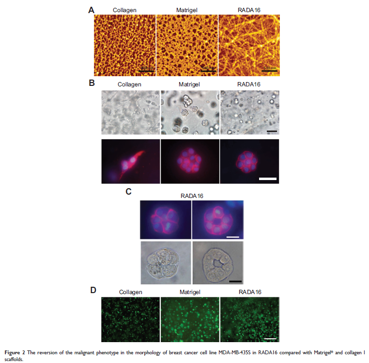
line MDA-MB-435S contained a high percentage (>99%) of CD44+/CD24- cells, which exhibited phenotypic reversion in 3D RADA16 nanofiber scaffold
compared with collagen I and Matrigel. The newly formed reverted acini-like
colonies reassembled a basement membrane and reorganized their cytoskeletons.
At the same time, cells cultured and embedded in RADA16 peptide scaffold
exhibited growth arrest. Also, they exhibited different migration potential,
which links their migration ability with their cellular morphology. Consistent
with studies in vitro, the in vivo tumor formation assay further supported of
the functional changes caused by the reversion in 3D RADA16 culture. Expression
levels of intercellular surface adhesion molecule-1 were upregulated in cells
cultured in RADA16 scaffolds, and the NF-kappa B inhibitor pyrrolidine
dithiocarbamate could inhibit RADA16-induced upregulation of intercellular
surface adhesion molecule-1 and the phenotype reversion of MDA-MB-453S cells.
Conclusion: Culturing a CD44+/CD24--enriched
breast cancer cell population in 3D RADA16 peptide nanofiber scaffold led to a
significant phenotypic reversion compared with Matrigel and collagen I.
Keywords: 3D culture,
phenotype, reversion
