108384
论文已发表
注册即可获取德孚的最新动态
IF 收录期刊
- 3.4 Breast Cancer (Dove Med Press)
- 3.2 Clin Epidemiol
- 2.6 Cancer Manag Res
- 2.9 Infect Drug Resist
- 3.7 Clin Interv Aging
- 5.1 Drug Des Dev Ther
- 3.1 Int J Chronic Obstr
- 6.6 Int J Nanomed
- 2.6 Int J Women's Health
- 2.9 Neuropsych Dis Treat
- 2.8 OncoTargets Ther
- 2.0 Patient Prefer Adher
- 2.2 Ther Clin Risk Manag
- 2.5 J Pain Res
- 3.0 Diabet Metab Synd Ob
- 3.2 Psychol Res Behav Ma
- 3.4 Nat Sci Sleep
- 1.8 Pharmgenomics Pers Med
- 2.0 Risk Manag Healthc Policy
- 4.1 J Inflamm Res
- 2.0 Int J Gen Med
- 3.4 J Hepatocell Carcinoma
- 3.0 J Asthma Allergy
- 2.2 Clin Cosmet Investig Dermatol
- 2.4 J Multidiscip Healthc

OCT4,SOX2 和 NAN OG 阳性表达与 HER2 阳性乳腺癌患者的分化程度低、疾病进展和总体生存率较差相关
Authors Yang F, Zhang J, Yang H
Received 8 May 2018
Accepted for publication 24 August 2018
Published 6 November 2018 Volume 2018:11 Pages 7873—7881
DOI https://doi.org/10.2147/OTT.S173522
Checked for plagiarism Yes
Review by Single-blind
Peer reviewers approved by Dr Cristina Weinberg
Peer reviewer comments 2
Editor who approved publication: Dr William Cho
Objective: This study aimed to evaluate the correlations of expression of
OCT4, SOX2, and NANOG with clinicopathological features and overall survival
(OS) in human epidermal growth factor receptor 2-positive (HER2+) breast cancer (BC) patients.
Methods: One hundred and thirty-four surgical HER2+ BC patients who
received doxorubicin and cyclophosphamide followed by paclitaxel and trastuzumab
adjuvant therapy were enrolled in this study. Immunofluorescence assay was used
to detect OCT4, SOX2, and NANOG expressions. The median follow-up duration was
104 months, and the last follow-up date was December 31, 2017.
Results: The expressions of OCT4 (P =0.001), SOX2 (P =0.003), and NANOG (P =0.005) were higher in tumor
tissues compared with paired adjacent tissues. OCT4 positive expression was
associated with poor pathological differentiation (P =0.028),
larger tumor size (P =0.022), advanced
N stage (P <0.001), and higher TNM stage
(P <0.001). SOX2 positive
expression was correlated with poor pathological differentiation (P =0.005), larger tumor size (P =0.013), and increased T stage (P =0.024). NANOG positive
expression was associated with poor pathological differentiation (P =0.028), higher N stage (P =0.001), and elevated TNM stage (P =0.001). Kaplan–Meier curves
disclosed that OCT4 (P =0.001) and NANOG
(P =0.001) positive expressions were
associated with worse OS, while SOX2 (P =0.058) positive
expression was only numerically correlated with poor OS, but without
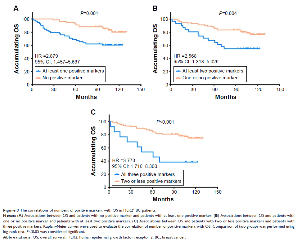
statistical significance. Further analyses revealed that co-expression of these
three biomarkers disclosed even better predictive value for shorter OS.
Conclusion: OCT4, SOX2, and NANOG positive expressions correlate with poor
differentiation and advanced disease stage, and OCT4 and NANOG present with
predictive values for poor OS in HER2+BC patients.
Keywords: clinicopathological features, prognosis, biomarker, tumor tissue,
predictive value
