108605
论文已发表
注册即可获取德孚的最新动态
IF 收录期刊
- 3.4 Breast Cancer (Dove Med Press)
- 3.2 Clin Epidemiol
- 2.6 Cancer Manag Res
- 2.9 Infect Drug Resist
- 3.7 Clin Interv Aging
- 5.1 Drug Des Dev Ther
- 3.1 Int J Chronic Obstr
- 6.6 Int J Nanomed
- 2.6 Int J Women's Health
- 2.9 Neuropsych Dis Treat
- 2.8 OncoTargets Ther
- 2.0 Patient Prefer Adher
- 2.2 Ther Clin Risk Manag
- 2.5 J Pain Res
- 3.0 Diabet Metab Synd Ob
- 3.2 Psychol Res Behav Ma
- 3.4 Nat Sci Sleep
- 1.8 Pharmgenomics Pers Med
- 2.0 Risk Manag Healthc Policy
- 4.1 J Inflamm Res
- 2.0 Int J Gen Med
- 3.4 J Hepatocell Carcinoma
- 3.0 J Asthma Allergy
- 2.2 Clin Cosmet Investig Dermatol
- 2.4 J Multidiscip Healthc

MnO 纳米粒子具有用于磁共振成像和心肌梗死药物输送的潜在应用
Authors Zheng Y, Zhang H, Hu Y, Bai L, Xue J
Received 5 June 2018
Accepted for publication 22 August 2018
Published 8 October 2018 Volume 2018:13 Pages 6177—6188
DOI https://doi.org/10.2147/IJN.S176404
Checked for plagiarism Yes
Review by Single-blind
Peer reviewers approved by Dr Alexander Kharlamov
Peer reviewer comments 2
Editor who approved publication: Dr Lei Yang
Background: Myocardial infarction (MI) is a leading cause of death worldwide. Therefore, nanoparticles that applied for specific diagnosis of the infarcted area and/or local myocardial delivery of therapeutic agents, are highly desired.
Materials and methods: Herein, we developed the MnO-based nanoparticles, with magnetic resonance (MR) and near-infrared fluorescence imaging modalities as an MR imaging contrast agent and potential drug vehicle for the detection and treatment of MI. The chemophysical characteristics, targeting ability toward infarcted myocardium, biodistribution, and biocompatibility of the MnO-based nanoparticles were studied.
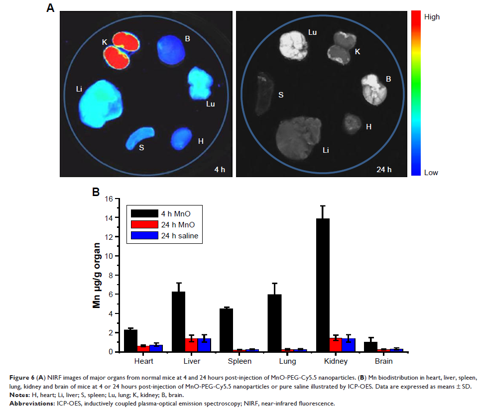
Results: It was found that the MnO-based dual-modal nanoparticles possess high r 1 relaxivity and induced no notable in vitro or in vivo toxicity. In a rat model of MI, these nanoparticles represent a very promising MR imaging contrast agent for sensitive and specific detection of the infarcted area, more importantly, without cardiotoxicity, the major defect of conventional Mn-based contrasts. Moreover, ex vivo near-infrared fluorescence imaging indicated that the MnO nanoparticles preferentially accumulate in the infarcted myocardium, which makes them an ideal drug vehicle for MI treatment.
Conclusion: In summary, the use of these MnO nanoparticles as a T1-weighted MR imaging contrast agent and potential drug vehicle to target the infarcted myocardium may provide new opportunities for accurate detection of myocardial infarct and treatment of ischemic heart diseases.
Keywords: Cy5.5 conjugated MnO nanoparticles, Dual-modal nanoprobe, Near-infrared fluorescence imaging, Myocardial ischemia
