108552
论文已发表
注册即可获取德孚的最新动态
IF 收录期刊
- 3.4 Breast Cancer (Dove Med Press)
- 3.2 Clin Epidemiol
- 2.6 Cancer Manag Res
- 2.9 Infect Drug Resist
- 3.7 Clin Interv Aging
- 5.1 Drug Des Dev Ther
- 3.1 Int J Chronic Obstr
- 6.6 Int J Nanomed
- 2.6 Int J Women's Health
- 2.9 Neuropsych Dis Treat
- 2.8 OncoTargets Ther
- 2.0 Patient Prefer Adher
- 2.2 Ther Clin Risk Manag
- 2.5 J Pain Res
- 3.0 Diabet Metab Synd Ob
- 3.2 Psychol Res Behav Ma
- 3.4 Nat Sci Sleep
- 1.8 Pharmgenomics Pers Med
- 2.0 Risk Manag Healthc Policy
- 4.1 J Inflamm Res
- 2.0 Int J Gen Med
- 3.4 J Hepatocell Carcinoma
- 3.0 J Asthma Allergy
- 2.2 Clin Cosmet Investig Dermatol
- 2.4 J Multidiscip Healthc

肿瘤血管正常化的监测:从基础研究到临床应用的关键点
Authors Li W, Quan Y, Li Y, Lu L, Cui M
Received 19 May 2018
Accepted for publication 13 July 2018
Published 3 October 2018 Volume 2018:10 Pages 4163—4172
DOI https://doi.org/10.2147/CMAR.S174712
Checked for plagiarism Yes
Review by Single-blind
Peer reviewers approved by Dr Colin Mak
Peer reviewer comments 2
Editor who approved publication: Professor Nakshatri
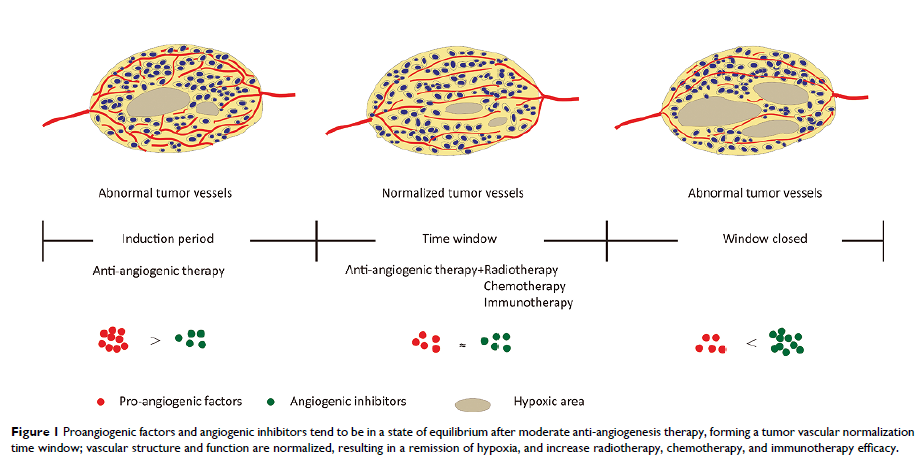
Abstract: Tumor vascular normalization alleviates hypoxia in the tumor
microenvironment, reduces the degree of malignancy, and increases the efficacy
of traditional therapy. However, the time window for vascular normalization is
narrow; therefore, how to determine the initial and final points of the time
window accurately is a key factor in combination therapy. At present, the gold
standard for detecting the normalization of tumor blood vessels is histological
staining, including tumor perfusion, microvessel density (MVD), vascular
morphology, and permeability. However, this detection method is almost
unrepeatable in the same individual and does not dynamically monitor the trend
of the time window; therefore, finding a relatively simple and specific
monitoring index has important clinical significance. Imaging has long been
used to assess changes in tumor blood vessels and tumor changes caused by the
oxygen environment in clinical practice; some preclinical and clinical research
studies demonstrate the feasibility to assess vascular changes, and some new
methods were in preclinical research. In this review, we update the most recent
insights of evaluating tumor vascular normalization.
Keywords: angiogenesis, vascular normalization, time window
