108384
论文已发表
注册即可获取德孚的最新动态
IF 收录期刊
- 3.4 Breast Cancer (Dove Med Press)
- 3.2 Clin Epidemiol
- 2.6 Cancer Manag Res
- 2.9 Infect Drug Resist
- 3.7 Clin Interv Aging
- 5.1 Drug Des Dev Ther
- 3.1 Int J Chronic Obstr
- 6.6 Int J Nanomed
- 2.6 Int J Women's Health
- 2.9 Neuropsych Dis Treat
- 2.8 OncoTargets Ther
- 2.0 Patient Prefer Adher
- 2.2 Ther Clin Risk Manag
- 2.5 J Pain Res
- 3.0 Diabet Metab Synd Ob
- 3.2 Psychol Res Behav Ma
- 3.4 Nat Sci Sleep
- 1.8 Pharmgenomics Pers Med
- 2.0 Risk Manag Healthc Policy
- 4.1 J Inflamm Res
- 2.0 Int J Gen Med
- 3.4 J Hepatocell Carcinoma
- 3.0 J Asthma Allergy
- 2.2 Clin Cosmet Investig Dermatol
- 2.4 J Multidiscip Healthc

米非司酮通过调节 FAK 和 PI3K/AKT 信号通路抑制 HUUA 细胞的增殖、迁移和侵袭,促进其凋亡
Authors Sang L, Lu D, Zhang J, Du S, Zhao X
Received 2 April 2018
Accepted for publication 4 July 2018
Published 4 September 2018 Volume 2018:11 Pages 5441—5449
DOI https://doi.org/10.2147/OTT.S169947
Checked for plagiarism Yes
Review by Single-blind
Peer reviewers approved by Dr Cristina Weinberg
Peer reviewer comments 2
Editor who approved publication: Dr XuYu Yang
Purpose: The aim was to investigate mifepristone effects on endometrial carcinoma and the related mechanism.
Methods: HHUA cells were treated with DMEM containing different concentrations of mifepristone. HHUA cells treated with 100 µmol/L mifepristone were named the Mifepristone group. HHUA cells co-transfected with pcDNA3.1-PI3K and pcDNA3.1-AKT overexpression vectors were treated with 100 µmol/L mifepristone and named the Mifepristone + PI3K/AKT group. mRNA expression was detected by quantitative reverse transcription PCR. Protein expression was performed by Western blot. Cell proliferation was conducted by MTT assay. Wound-healing assay was conducted. Transwell was used to detect cells migration and invasion. Apoptosis detection was performed by flow cytometry.
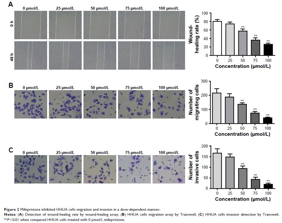
Results: Mifepristone inhibited HHUA cells proliferation in a dose-dependent manner. Compared with HHUA cells treated with 0 µmol/L mifepristone, HHUA cells treated by 50–100 µmol/L mifepristone had a lower wound-healing rate, a greater number of migrating and invasive cells (P <0.01), as well as a higher percentage of apoptotic cells and Caspase-3 expression (P <0.01). When HHUA cells were treated with 50–100 µmol/L of mifepristone, FAK, p-FAK, p-PI3K and p-AKT relative expression was all significantly lower than HHUA cells treated with 0 µmol/L of mifepristone (P <0.01). Compared with the Mifepristone group, HHUA cells of the Mifepristone + PI3K/AKT group had a lower cell growth inhibition rate and percentage of apoptotic cells (P <0.01).
Conclusion: Mifepristone inhibited HUUA cells proliferation, migration and invasion and promoted its apoptosis by regulation of FAK and PI3K/AKT signaling pathway.
Keywords: Mifepristone, HHUA cells, proliferation, FAK, PI3K/AKT signaling pathway
