108384
论文已发表
注册即可获取德孚的最新动态
IF 收录期刊
- 3.4 Breast Cancer (Dove Med Press)
- 3.2 Clin Epidemiol
- 2.6 Cancer Manag Res
- 2.9 Infect Drug Resist
- 3.7 Clin Interv Aging
- 5.1 Drug Des Dev Ther
- 3.1 Int J Chronic Obstr
- 6.6 Int J Nanomed
- 2.6 Int J Women's Health
- 2.9 Neuropsych Dis Treat
- 2.8 OncoTargets Ther
- 2.0 Patient Prefer Adher
- 2.2 Ther Clin Risk Manag
- 2.5 J Pain Res
- 3.0 Diabet Metab Synd Ob
- 3.2 Psychol Res Behav Ma
- 3.4 Nat Sci Sleep
- 1.8 Pharmgenomics Pers Med
- 2.0 Risk Manag Healthc Policy
- 4.1 J Inflamm Res
- 2.0 Int J Gen Med
- 3.4 J Hepatocell Carcinoma
- 3.0 J Asthma Allergy
- 2.2 Clin Cosmet Investig Dermatol
- 2.4 J Multidiscip Healthc

具有终末期肾病的神经性无症状患者的异常皮质厚度
Authors Dong J, Ma X, Lin W, Liu M, Fu S, Yang L, Jiang G
Received 4 April 2018
Accepted for publication 24 May 2018
Published 2 August 2018 Volume 2018:14 Pages 1929—1939
DOI https://doi.org/10.2147/NDT.S170106
Checked for plagiarism Yes
Review by Single-blind
Peer reviewers approved by Prof. Dr. Roumen Kirov
Peer reviewer comments 3
Editor who approved publication: Professor Wai Kwong Tang
Purpose: The aim of this study is to investigate the morphology of cortical
gray matter in patients with end-stage renal disease (ESRD) and the
relationship between cortical thickness and kidney function.
Patients and
methods: Three-dimensional high-resolution
brain structural magnetic resonance imaging data were collected from 35
patients with ESRD (28 men, 18–61 years old) and 40 age- and gender-matched
healthy controls (HCs, 32 men, 22–58 years old). Vertex-wise analysis was then
performed to compare the brains of the patients with ESRD with those of HCs to
identify abnormalities in the brains of the former. Multiple biochemical
measures of renal metabolin, vascular risk factors, general cognitive ability,
and dialysis duration were correlated with brain morphometry alterations for
the patients.
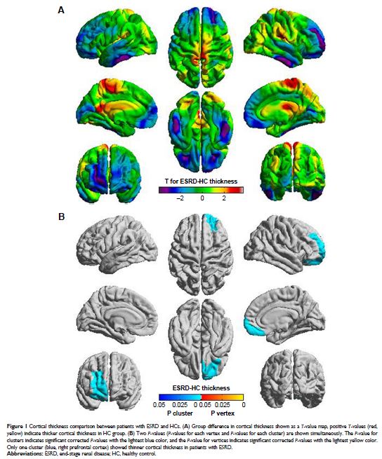
Results: Patients with ESRD showed lesser cortical thickness than the HCs.
The most significant cluster with decreased cortical thickness was found in the
right prefrontal cortex (P <0.05,
random-field theory correction). In addition, the four local peak vertices in
the prefrontal cluster were lateral prefrontal cortex (Peaks 1 and 2), medial
prefrontal cortex (Peak 3), and ventral prefrontal cortex (Peak 4). Significant
negative correlations were observed between the cortical thicknesses of all
four peak vertices and blood urea nitrogen; a negative correlation, between the
cortical thickness in three of four peaks and serum creatinine; and a positive
correlation, between cortical thickness in the medial prefrontal cortex (Peak
3) and hemoglobin.
Conclusion: These results provided compelling evidence for cortical
abnormality of ESRD patients and suggested that kidney function may be the key
factor for predicting changes of brain tissue structure.
Keywords: cortical thickness, kidney function, vertex-wise analysis,
multivariate analysis, brain morphometry alterations
