108605
论文已发表
注册即可获取德孚的最新动态
IF 收录期刊
- 3.4 Breast Cancer (Dove Med Press)
- 3.2 Clin Epidemiol
- 2.6 Cancer Manag Res
- 2.9 Infect Drug Resist
- 3.7 Clin Interv Aging
- 5.1 Drug Des Dev Ther
- 3.1 Int J Chronic Obstr
- 6.6 Int J Nanomed
- 2.6 Int J Women's Health
- 2.9 Neuropsych Dis Treat
- 2.8 OncoTargets Ther
- 2.0 Patient Prefer Adher
- 2.2 Ther Clin Risk Manag
- 2.5 J Pain Res
- 3.0 Diabet Metab Synd Ob
- 3.2 Psychol Res Behav Ma
- 3.4 Nat Sci Sleep
- 1.8 Pharmgenomics Pers Med
- 2.0 Risk Manag Healthc Policy
- 4.1 J Inflamm Res
- 2.0 Int J Gen Med
- 3.4 J Hepatocell Carcinoma
- 3.0 J Asthma Allergy
- 2.2 Clin Cosmet Investig Dermatol
- 2.4 J Multidiscip Healthc

羟考酮可改善原代小胶质细胞中脂多糖诱导的炎症反应
Authors Ye J, Yan H, Xia Z
Received 22 December 2017
Accepted for publication 3 April 2018
Published 22 June 2018 Volume 2018:11 Pages 1199—1207
DOI https://doi.org/10.2147/JPR.S160659
Checked for plagiarism Yes
Review by Single-blind
Peer reviewers approved by Dr Amy Norman
Peer reviewer comments 2
Editor who approved publication: Dr Katherine Hanlon
Background: Activation
of microglia participates in a wide range of pathophysiological processes in
the central nervous system. Some studies reported that oxycodone
(6-deoxy-7,8-dehydro-14-hydroxy-3-O-methyl-6oxomorphine) could inhibit the
overactivation of glial cells in rats’ spinal cords. In the present study, we
observed the effect of oxycodone on inflammatory molecules and pathway in
lipopolysaccharide (LPS)-stimulated primary microglia in rats.
Materials and
methods: Neonatal rats’ primary microglia
were exposed to various concentrations (25, 50, 100 ng/mL) of oxycodone for 1 h
after LPS stimulation for 24 h. The levels of pro-inflammatory mediators,
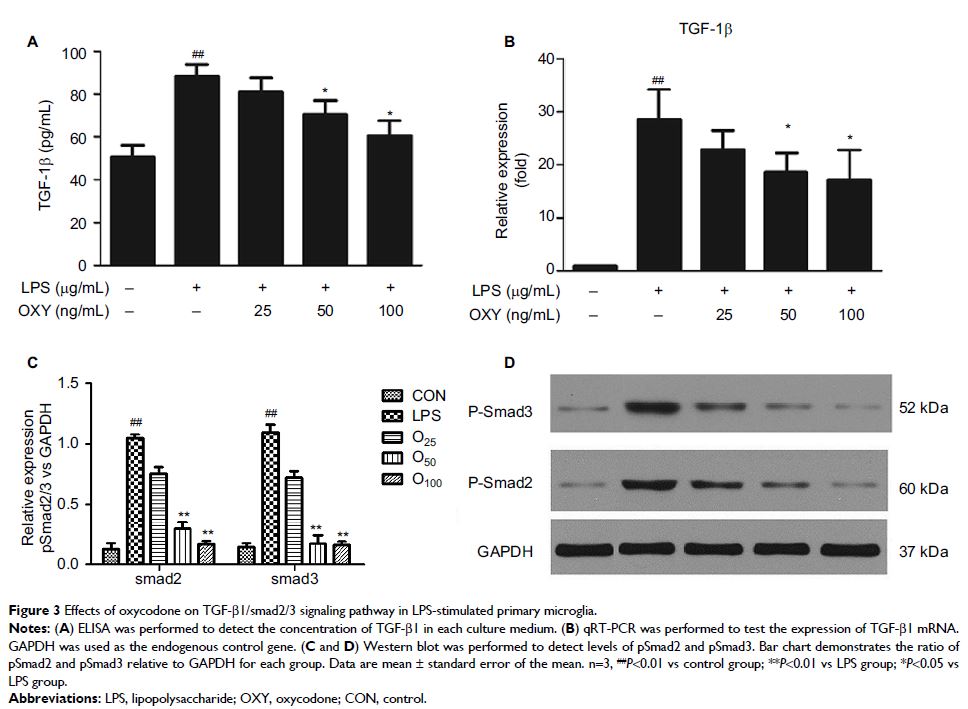
IL-1β, TNF-α, and TGF-β1/smad2/3 signaling pathway were measured. The
activation situation of microglia and the expression of TβR1 were observed by
immunofluorescence.
Results: Oxycodone at 25 ng/mL did not change the levels of proinflammatory
molecules and TGF-β1/smad2/3 signaling pathway in primary microglia, which was
increased by LPS. Oxycodone at 50 and 100 ng/mL could significantly suppress
LPS-induced production of TNF-α and IL-1β and the expression of TNF-αmRNA,
IL-1βmRNA, and TGF-β1/smad2/3 signaling pathway.
Conclusion: These findings indicate that oxycodone, at relatively high
clinically relevant concentration, can inhibit inflammatory response in
LPS-induced primary microglia. The detailed mechanism needs to be investigated
in future.
Keywords: oxycodone, inflammatory, microglia
