108985
论文已发表
注册即可获取德孚的最新动态
IF 收录期刊
- 3.4 Breast Cancer (Dove Med Press)
- 3.2 Clin Epidemiol
- 2.6 Cancer Manag Res
- 2.9 Infect Drug Resist
- 3.7 Clin Interv Aging
- 5.1 Drug Des Dev Ther
- 3.1 Int J Chronic Obstr
- 6.6 Int J Nanomed
- 2.6 Int J Women's Health
- 2.9 Neuropsych Dis Treat
- 2.8 OncoTargets Ther
- 2.0 Patient Prefer Adher
- 2.2 Ther Clin Risk Manag
- 2.5 J Pain Res
- 3.0 Diabet Metab Synd Ob
- 3.2 Psychol Res Behav Ma
- 3.4 Nat Sci Sleep
- 1.8 Pharmgenomics Pers Med
- 2.0 Risk Manag Healthc Policy
- 4.1 J Inflamm Res
- 2.0 Int J Gen Med
- 3.4 J Hepatocell Carcinoma
- 3.0 J Asthma Allergy
- 2.2 Clin Cosmet Investig Dermatol
- 2.4 J Multidiscip Healthc

PRDM 基因启动子甲基化在非小细胞肺癌中的作用
Authors Tan SX, Hu RC, Xia Q, Tan YL, Liu JJ, Gan GX, Wang LL
Received 11 November 2017
Accepted for publication 25 March 2018
Published 22 May 2018 Volume 2018:11 Pages 2991—3002
DOI https://doi.org/10.2147/OTT.S156775
Checked for plagiarism Yes
Review by Single-blind
Peer reviewers approved by Dr Justinn Cochran
Peer reviewer comments 4
Editor who approved publication: Dr Jianmin Xu
Background: Non–small
cell lung cancer (NSCLC) is one of the leading malignant tumors worldwide.
Aberrant gene promoter methylation contributes to NSCLC, and PRDM is a tumor
suppressor gene family that possesses histone methyltransferase activity. This
study aimed to investigate whether aberrant methylation of PRDM promoter is
involved in NSCLC.
Materials and
methods: Primary tumor tissues, adjacent
nontumorous tissues, and distant lung tissues were collected from 75 NSCLC
patients including 52 lung squamous cell carcinoma (LSCC) patients and 23 lung
adenocarcinoma patients. The expression of PRDMs was detected by polymerase
chain reaction (PCR), Western blot, and immunohistochemical analysis. The
methylation of PRDM promoters was detected by methylation-specific PCR. The
correlation of methylation and expression of PRDMs with clinicopathological
characteristics of patients were analyzed.
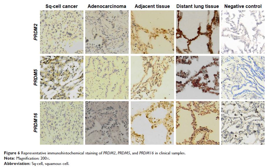
Results: mRNA expression of PRDM2 , PRDM5 , and PRDM16 was low or absent in
tumor tissues compared to distant lung tissues. The methylation frequencies
of PRDM2 , PRDM5 , and PRDM16 in tumor tissues were
significantly higher than those in distal lung tissues. In LSCC patients,
methylation of PRDM2 and PRDM16 was correlated with
smoking status and methylation of PRDM5 was correlated with tumor
differentiation.
Conclusion: The expression of PRDM2 , PRDM5 , and PRDM16 is low or absent in
NSCLC, and this is mainly due to gene promoter methylation. Smoking may be an
important cause of PRDM2 and PRDM16 methylation in NSCLC.
Keywords: lung tumor, PR domain zinc finger protein, epigenetics,
methylation, gene expression
