108985
论文已发表
注册即可获取德孚的最新动态
IF 收录期刊
- 3.4 Breast Cancer (Dove Med Press)
- 3.2 Clin Epidemiol
- 2.6 Cancer Manag Res
- 2.9 Infect Drug Resist
- 3.7 Clin Interv Aging
- 5.1 Drug Des Dev Ther
- 3.1 Int J Chronic Obstr
- 6.6 Int J Nanomed
- 2.6 Int J Women's Health
- 2.9 Neuropsych Dis Treat
- 2.8 OncoTargets Ther
- 2.0 Patient Prefer Adher
- 2.2 Ther Clin Risk Manag
- 2.5 J Pain Res
- 3.0 Diabet Metab Synd Ob
- 3.2 Psychol Res Behav Ma
- 3.4 Nat Sci Sleep
- 1.8 Pharmgenomics Pers Med
- 2.0 Risk Manag Healthc Policy
- 4.1 J Inflamm Res
- 2.0 Int J Gen Med
- 3.4 J Hepatocell Carcinoma
- 3.0 J Asthma Allergy
- 2.2 Clin Cosmet Investig Dermatol
- 2.4 J Multidiscip Healthc

分层排列的纤维蛋白纳米纤维水凝胶可加速轴突再生和大鼠脊髓损伤的运动功能恢复
Authors Yao S, Yu S, Cao Z, Yang Y, Yu X, Mao HQ, Wang LN, Sun X, Zhao L, Wang XM
Received 8 December 2017
Accepted for publication 28 February 2018
Published 17 May 2018 Volume 2018:13 Pages 2883—2895
DOI https://doi.org/10.2147/IJN.S159356
Checked for plagiarism Yes
Review by Single-blind
Peer reviewers approved by Dr Cristina Weinberg
Peer reviewer comments 2
Editor who approved publication: Dr Linlin Sun
Background: Designing novel biomaterials that incorporate or mimic the functions of
extracellular matrix to deliver precise regulatory signals for tissue
regeneration is the focus of current intensive research efforts in tissue
engineering and regenerative medicine.
Methods and
results: To mimic the natural
environment of the spinal cord tissue, a three-dimensional hierarchically
aligned fibrin hydrogel (AFG) with oriented topography and soft stiffness has
been fabricated by electrospinning and a concurrent molecular self-assembling
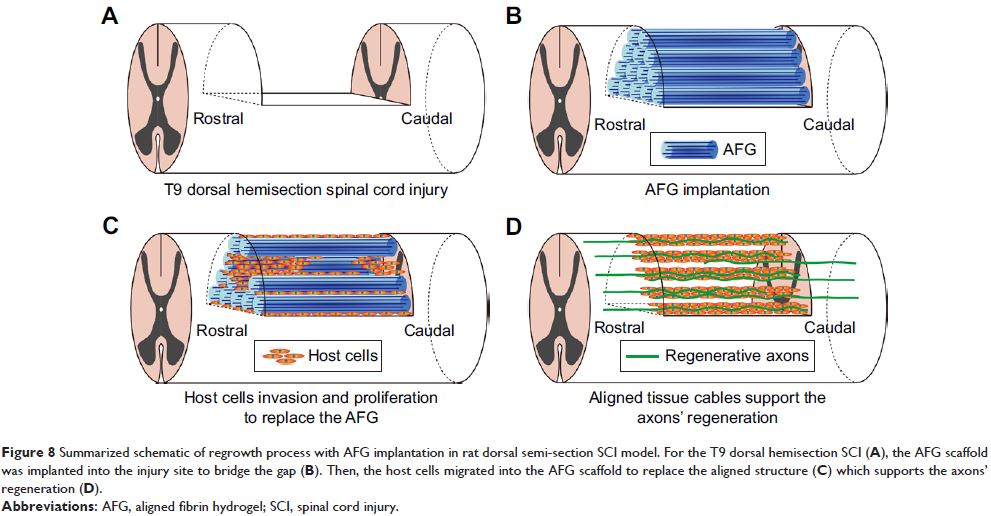
process. In this study, the AFG was implanted into a rat dorsal hemisected
spinal cord injury model to bridge the lesion site. Host cells invaded promptly
along the aligned fibrin hydrogels to form aligned tissue cables in the first
week, and then were followed by axonal regrowth. At 4 weeks after the surgery,
neurofilament (NF)-positive staining fibers were detected near the rostral end
as well as the middle site of defect, which aligned along the tissue cables.
Abundant NF- and GAP-43-positive staining indicated new axon regrowth in the
oriented tissue cables, which penetrated throughout the lesion site in 8 weeks.
Additionally, the abundant blood vessels marked with RECA-1 had reconstructed
within the lesion site at 4 weeks after surgery. Basso-Beattie-Bresnahan
scoring showed that the locomotor performance of the AFG group recovered much
faster than that of blank control group or the random fibrin hydrogel (RFG) group
from 2 weeks after surgery. Furthermore, diffusion tensor imaging tractography
of MRI confirmed the optimal axon fiber reconstruction compared with the RFG
and control groups.
Conclusion: Taken together, our results suggested that the AFG scaffold provided an
inductive matrix for accelerating directional host cell invasion, vascular
system reconstruction, and axonal regrowth, which could promote and support
extensive aligned axonal regrowth and locomotor function recovery.
Keywords: spinal cord injury, nerve regrowth, fibrin hydrogel, aligned
structure, soft stiffness
