116231
论文已发表
注册即可获取德孚的最新动态
IF 收录期刊
- 3.6 Breast Cancer (Dove Med Press)
- 4.3 Clin Epidemiol
- 2.6 Cancer Manag Res
- 3.2 Infect Drug Resist
- 4.1 Clin Interv Aging
- 6.1 Drug Des Dev Ther
- 4.1 Int J Chronic Obstr
- 8.7 Int J Nanomed
- 2.5 Int J Women's Health
- 3.2 Neuropsych Dis Treat
- 2.4 OncoTargets Ther
- 2.6 Patient Prefer Adher
- 2.6 Ther Clin Risk Manag
- 3.1 J Pain Res
- 3.5 Diabet Metab Synd Ob
- 4.5 Psychol Res Behav Ma
- 3.4 Nat Sci Sleep
- 2.4 Pharmgenomics Pers Med
- 2.6 Risk Manag Healthc Policy
- 4.6 J Inflamm Res
- 2.3 Int J Gen Med
- 3.9 J Hepatocell Carcinoma
- 3.3 J Asthma Allergy
- 2.5 Clin Cosmet Investig Dermatol
- 3.0 J Multidiscip Healthc

RADA16-I 纳米纤维的淀粉样蛋白染色性能及其在纳米材料检测和成像中的潜在应用
Authors Chen Y, Hua Y, Zhang W, Tang C, Wang Y, Zhang Y, Qiu F
Received 14 December 2017
Accepted for publication 17 February 2018
Published 23 April 2018 Volume 2018:13 Pages 2477—2489
DOI https://doi.org/10.2147/IJN.S159785
Checked for plagiarism Yes
Review by Single-blind
Peer reviewers approved by Dr Mohankandhasamy Ramasamy
Peer reviewer comments 3
Editor who approved publication: Dr Linlin Sun
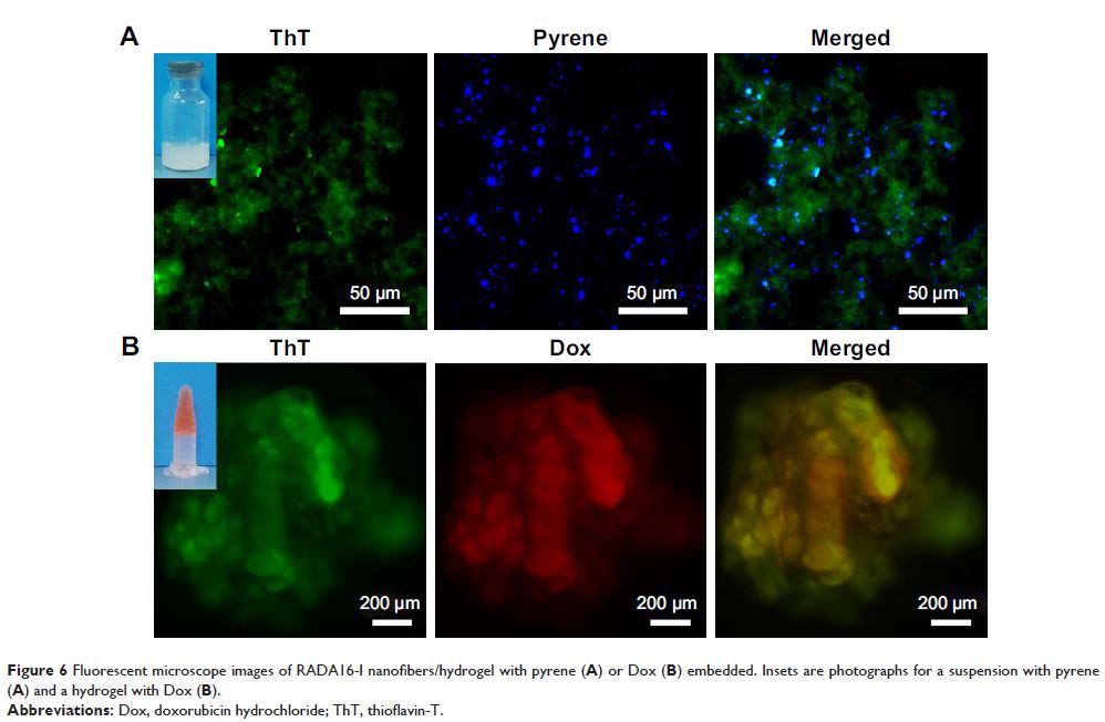
Background: Designer
self-assembling peptide nanofibers (SAPNFs) as a novel kind of emerging
nanomaterial have received more and more attention in the field of nanomedicine
in recent years. However, a simple method to monitor and image SAPNFs is still
currently absent.
Methods: RADA16-I, a well-studied ionic complementary peptide was used as a
model to check potential amyloid-like staining properties of SAPNFs.
Thioflavin-T (ThT) and Congo red (CR) as specific dyes for amyloid-like fibrils
were used to stain RADA16-I nanofibers in solution, combined with drugs or
cells, or injected in vivo as hydrogels. Fluorescent spectrometry and
fluorescent microscopy were used to check ThT-binding property, and polarized
light microscopy was used to check CR-staining property.
Results: ThT binding with the nanofibers showed enhanced and blue-shifted
fluorescence, and specific apple-green birefringence could be observed after
the nanofibers were stained with CR. Based on these properties we further
showed that ThT-binding fluorescence intensity could be used to monitor the
forming and changing of nanofibers in solution, while fluorescent microscopy
and polarized light microscopy could be used to image the nanofibers as
material for drug delivery, 3D cell culture, and tissue regeneration.
Conclusion: Our results may provide convenient and reliable tools for
detecting SAPNFs, which would be helpful for understanding their
self-assembling process and exploring their applications.
Keywords: self-assembling peptides, nanofibers, amyloid fibrils,
thioflavin-T, Congo red