116231
论文已发表
注册即可获取德孚的最新动态
IF 收录期刊
- 3.6 Breast Cancer (Dove Med Press)
- 4.3 Clin Epidemiol
- 2.6 Cancer Manag Res
- 3.2 Infect Drug Resist
- 4.1 Clin Interv Aging
- 6.1 Drug Des Dev Ther
- 4.1 Int J Chronic Obstr
- 8.7 Int J Nanomed
- 2.5 Int J Women's Health
- 3.2 Neuropsych Dis Treat
- 2.4 OncoTargets Ther
- 2.6 Patient Prefer Adher
- 2.6 Ther Clin Risk Manag
- 3.1 J Pain Res
- 3.5 Diabet Metab Synd Ob
- 4.5 Psychol Res Behav Ma
- 3.4 Nat Sci Sleep
- 2.4 Pharmgenomics Pers Med
- 2.6 Risk Manag Healthc Policy
- 4.6 J Inflamm Res
- 2.3 Int J Gen Med
- 3.9 J Hepatocell Carcinoma
- 3.3 J Asthma Allergy
- 2.5 Clin Cosmet Investig Dermatol
- 3.0 J Multidiscip Healthc

使用多模式成像剂无创评估移植后的内皮祖细胞在局部缺血肌肉中的迁移效应
Authors Peng XG, Li C, Bai Y, Wang X, Zhang Y, An Y, Teng GJ, Ju S
Received 30 September 2017
Accepted for publication 7 December 2017
Published 22 March 2018 Volume 2018:13 Pages 1819—1829
DOI https://doi.org/10.2147/IJN.S152976
Checked for plagiarism Yes
Review by Single-blind
Peer reviewers approved by Dr Govarthanan Muthusamy
Peer reviewer comments 2
Editor who approved publication: Dr Linlin Sun
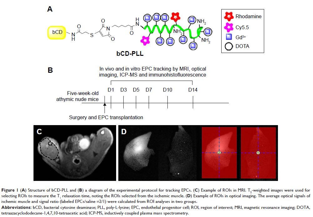
Background: Endothelial progenitor cells (EPCs) play an important role in
repairing ischemia tissues. However, the survival, migration and therapeutic
efficacy of EPCs after transplantation need to be better understood for further
cell therapy.
Purpose: This study investigated the migration effect of EPCs labeled with
a multimodal imaging agent in a murine ischemic hindlimb model, using magnetic
resonance imaging (MRI) and optical imaging after transplantation.
Methods: EPCs derived from mouse bone marrow were labeled with a multimodal
imaging agent and were administered through intracardiac delivery to mice with
ischemic hindlimbs. The injected EPCs and their migration effect were observed
via MRI and optical imaging in vivo, and then compared to a reference standard
based on histological data. The quantification of gadolinium in tissue samples
was done using inductively coupled plasma mass spectrometry (ICP-MS).
Results: Using in vivo MRI and optical imaging, the labeled EPCs were
observed to migrate to ischemic muscle on days 3–5 after injection, while ex
vivo, the EPCs were observed in the capillary vessels of the injured tissue.
There were significant linear correlations between the Gd contents measured
using ICP-MS in samples from the ischemic hindlimbs and livers and T1 relaxation times calculated using MRI, as well as the average
fluorescence signal intensities recorded in optical images (T1 relaxation time: r =0.491; average
signal from optical imaging: r =0.704, P <0.01). EPC treatment
upregulated the levels of C-X-C chemokine receptor 4 and vascular endothelial
growth factor (VEGF) receptor 2 and enhanced the expression of stromal
cell-derived factor-1 and VEGF.
Conclusion: Transplanted EPCs can be monitored with noninvasive MRI and
optical imaging in vivo and were found to enhance the paracrine secretion of
angiogenic factors.
Keywords: endothelial progenitor cell, ischemia, regeneration, cell
tracking, magnetic resonance imaging, optical imaging