116231
论文已发表
注册即可获取德孚的最新动态
IF 收录期刊
- 3.6 Breast Cancer (Dove Med Press)
- 4.3 Clin Epidemiol
- 2.6 Cancer Manag Res
- 3.2 Infect Drug Resist
- 4.1 Clin Interv Aging
- 6.1 Drug Des Dev Ther
- 4.1 Int J Chronic Obstr
- 8.7 Int J Nanomed
- 2.5 Int J Women's Health
- 3.2 Neuropsych Dis Treat
- 2.4 OncoTargets Ther
- 2.6 Patient Prefer Adher
- 2.6 Ther Clin Risk Manag
- 3.1 J Pain Res
- 3.5 Diabet Metab Synd Ob
- 4.5 Psychol Res Behav Ma
- 3.4 Nat Sci Sleep
- 2.4 Pharmgenomics Pers Med
- 2.6 Risk Manag Healthc Policy
- 4.6 J Inflamm Res
- 2.3 Int J Gen Med
- 3.9 J Hepatocell Carcinoma
- 3.3 J Asthma Allergy
- 2.5 Clin Cosmet Investig Dermatol
- 3.0 J Multidiscip Healthc

2 型糖尿病合并原发性肺结核患者 CT 征象与糖化血红蛋白水平的相关性
Authors Xia L, Li SF, Shao K, Zhang X, Huang S
Received 19 July 2017
Accepted for publication 19 October 2017
Published 30 January 2018 Volume 2018:11 Pages 187—193
DOI https://doi.org/10.2147/IDR.S146741
Checked for plagiarism Yes
Review by Single-blind
Peer reviewers approved by Dr Amy Norman
Peer reviewer comments 2
Editor who approved publication: Professor Suresh Antony
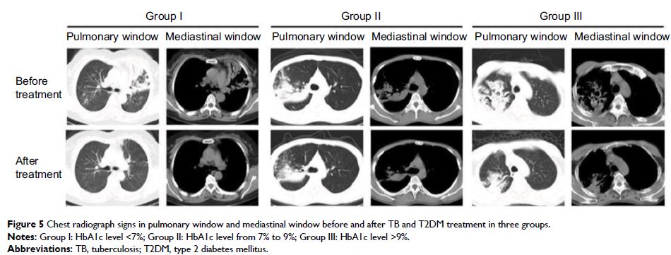
Abstract: To investigate the correlation between computed tomography (CT) features
and glycosylated hemoglobin (HbAlc) levels in patients with type 2 diabetes
mellitus (T2DM) complicated with primary pulmonary tuberculosis (PTB). One
hundred and eighty untreated PTB patients complicated with T2DM were selected.
Based on the HbAlc level, the patients were divided into three groups: HbAlc
level <7% (Group I: 32 patients), 7%–9% (Group II: 48 patients), and >9%
(Group III: 100 patients). The changes of CT manifestations and HbAlc were
analyzed after TB and T2DM treatment. In the three groups, the detection rate
of large segmented leafy shadow was 50%, 56.2%, and 87%; the air bronchogram
sign detection rate was 40.6%, 47.9%, and 77%; the discovery rate of
mouth-eaten cavity was 31.2%, 45.8%, and 65%; thick wall cavity detection rate
was 25%, 31.2%, and 52%; the rate of multiple cavities was 34.3%, 50%, and 73%;
and bronchial TB was found in 33.3%, 21.8%, and 46%, respectively. The
detection rates of lesions in Group III were significantly higher than in Group
II and Group I (p <0.05), and this increase was
significant (p <0.05). After treatment, the
HbAlc level reached control target (<7%) among all three groups and CT
absorption improvement rates were 100%, 72.9%, and 56% respectively. The
therapeutic efficacy of group I was better than group II (p <0.01), and the treatment
efficacy of group II was better than group III (p <0.05).
CT manifestations of T2DM complicated with PTB were closely related to HbAlc
level. The effect is better when HbAlc level <7%. HbAlc level effectively
reflects the severity and therapeutic effect to a certain extent. CT scan can
provide some important information for clinical imaging. The above two
examinations can guide clinicians to formulate the appropriate diagnosis and
treatment in a timely manner.
Keywords: T2DM, primary
pulmonary tuberculosis, CT, HbAlc