116231
论文已发表
注册即可获取德孚的最新动态
IF 收录期刊
- 3.6 Breast Cancer (Dove Med Press)
- 4.3 Clin Epidemiol
- 2.6 Cancer Manag Res
- 3.2 Infect Drug Resist
- 4.1 Clin Interv Aging
- 6.1 Drug Des Dev Ther
- 4.1 Int J Chronic Obstr
- 8.7 Int J Nanomed
- 2.5 Int J Women's Health
- 3.2 Neuropsych Dis Treat
- 2.4 OncoTargets Ther
- 2.6 Patient Prefer Adher
- 2.6 Ther Clin Risk Manag
- 3.1 J Pain Res
- 3.5 Diabet Metab Synd Ob
- 4.5 Psychol Res Behav Ma
- 3.4 Nat Sci Sleep
- 2.4 Pharmgenomics Pers Med
- 2.6 Risk Manag Healthc Policy
- 4.6 J Inflamm Res
- 2.3 Int J Gen Med
- 3.9 J Hepatocell Carcinoma
- 3.3 J Asthma Allergy
- 2.5 Clin Cosmet Investig Dermatol
- 3.0 J Multidiscip Healthc

在胸部肿瘤放疗的心脏周期中心脏、心包和左心室心肌运动的量化
Authors Tong Y, Yin Y, Lu J, Liu T, Chen J, Cheng P, Gong G
Received 31 October 2017
Accepted for publication 20 December 2017
Published 24 January 2018 Volume 2018:11 Pages 547—554
DOI https://doi.org/10.2147/OTT.S155680
Checked for plagiarism Yes
Review by Single-blind
Peer reviewers approved by Dr Ashok Kumar Pandurangan
Peer reviewer comments 3
Editor who approved publication: Dr Carlos Vigil Gonzales
Purpose: The purpose of this study was to quantify variations in the heart,
pericardium, and left ventricular myocardium (LVM) caused by cardiac movement
using the breath-hold technique.
Patients and
methods: In this study, the
electrocardiography-gated four-dimensional computed tomography (CT) images of 22
patients were analyzed, which were sorted into 20 phases (0–95%) according to
the cardiac cycle. The heart, pericardium, and LVM were contoured on each phase
of the CT images. The positions, volume, dice similarity coefficient (DSC) in
reference to 0% phase, and morphological parameters (max 3D diameter,
roundness, spherical disproportion, sphericity, and surface area) in different
phases of the heart, pericardium, and LVM were analyzed, which were presented
as mean ± standard deviation.
Results: The mean values of displacements along the X, Y, and Z axes
respectively were as follows: 1.2 mm, 0.6 mm, and 0.6 mm for the heart; 0.5 mm,
0.4 mm, and 0.8 mm for the pericardium; and 1.0 mm, 4.1 mm, and 1.9 mm for the
LVM. The maximum variations in volume and DSC respectively were 16.49%±3.85%
and 10.08%±2.14% for the heart, 12.62%±3.94% and 5.20%±1.54% for the
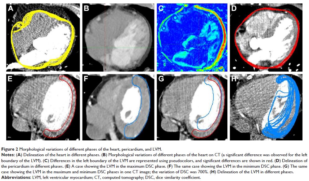
pericardium, and 24.23%±11.35% and 184.33%±128.61% for the LVM. The differences
in the morphological parameters between the maximum and minimum DSC phases for
the heart and pericardium were not significantly different (p >0.05) but were significantly
different for the LVM (p <0.05).
Conclusion: The volumetric and morphological variations of the heart were
similar to those of pericardium, and all were significantly smaller than those
of the LVM. This inconsistency in the volumetric and morphological variations
between the LVM and the heart and pericardium indicates that special protection
of the LVM should be considered.
Keywords: thoracic radiotherapy, cardiac activity, cardiac structures,
variations, volume, morphology