108985
论文已发表
注册即可获取德孚的最新动态
IF 收录期刊
- 3.4 Breast Cancer (Dove Med Press)
- 3.2 Clin Epidemiol
- 2.6 Cancer Manag Res
- 2.9 Infect Drug Resist
- 3.7 Clin Interv Aging
- 5.1 Drug Des Dev Ther
- 3.1 Int J Chronic Obstr
- 6.6 Int J Nanomed
- 2.6 Int J Women's Health
- 2.9 Neuropsych Dis Treat
- 2.8 OncoTargets Ther
- 2.0 Patient Prefer Adher
- 2.2 Ther Clin Risk Manag
- 2.5 J Pain Res
- 3.0 Diabet Metab Synd Ob
- 3.2 Psychol Res Behav Ma
- 3.4 Nat Sci Sleep
- 1.8 Pharmgenomics Pers Med
- 2.0 Risk Manag Healthc Policy
- 4.1 J Inflamm Res
- 2.0 Int J Gen Med
- 3.4 J Hepatocell Carcinoma
- 3.0 J Asthma Allergy
- 2.2 Clin Cosmet Investig Dermatol
- 2.4 J Multidiscip Healthc

柠檬酸二乙酯和柠檬酸钠降低纳米羟基磷灰石晶体对小鼠血管平滑肌细胞产生的细胞毒性作用
Authors Zhang CY, Sun XY, Ouyang JM, Gui BS
Received 3 July 2017
Accepted for publication 26 September 2017
Published 28 November 2017 Volume 2017:12 Pages 8511—8525
DOI https://doi.org/10.2147/IJN.S145386
Checked for plagiarism Yes
Review by Single-blind
Peer reviewers approved by Dr Govarthanan Muthusamy
Peer reviewer comments 2
Editor who approved publication: Dr Linlin Sun
Objective: This study aimed to investigate the damage mechanism of nanosized
hydroxyapatite (nano-HAp) on mouse aortic smooth muscle cells (MOVASs) and the
injury-inhibiting effects of diethyl citrate (Et2Cit) and sodium citrate (Na3Cit) to develop new drugs that can simultaneously induce anticoagulation
and inhibit vascular calcification.
Methods: The change in cell viability was evaluated using a cell
proliferation assay kit, and the amount of lactate dehydrogenase (LDH) released
was measured using an LDH kit. Intracellular reactive oxygen species (ROS) and
mitochondrial damage were detected by DCFH-DA staining and JC-1 staining. Cell
apoptosis and necrosis were detected by Annexin V staining. Intracellular
calcium concentration and lysosomal integrity were measured using Fluo-4/AM and
acridine orange, respectively.
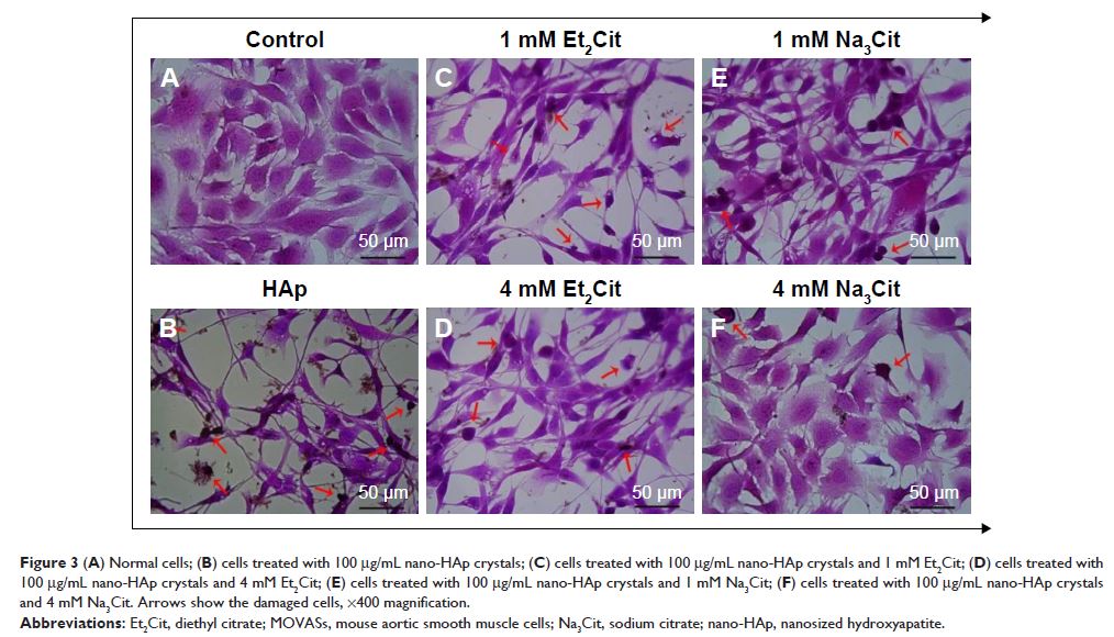
Results: Nano-HAp decreased cell viability and damaged the cell membrane,
resulting in the release of a large amount of LDH. Nano-HAp entered the cells
and damaged the mitochondria, and then induced cell apoptosis by producing a
large amount of ROS. In addition, nano-HAp increased the intracellular Ca2+ concentration, leading to lysosomal rupture and cell necrosis. On
addition of the anticoagulant Et2Cit or Na3Cit, cell viability and
mitochondrial membrane potential increased, whereas the amount of LDH released,
ROS, and apoptosis rate decreased. Et2Cit and Na3Cit could also chelate with Ca2+ to inhibit the intracellular Ca2+ elevations induced by nano-HAp, prevent lysosomal rupture, and
reduce cell necrosis. High concentrations of Et2Cit and Na3Cit exhibited strong inhibitory effects. The inhibitory capacity of Na3Cit was stronger than that of Et2Cit at similar concentrations.
Conclusion: Both Et2Cit and Na3Cit significantly reduced the cytotoxicity of nano-HAp on MOVASs and
inhibited the apoptosis and necrosis induced by nano-HAp crystals. The
chelating function of citrate resulted in both anticoagulation and binding to
HAp. Et2Cit and Na3Cit may play a role as anticoagulants in reducing injury to the vascular
wall caused by nano-HAp.
Keywords: vascular calcification, hydroxyapatite, diethyl citrate, sodium citrate,
cardiovascular disease
