116231
论文已发表
注册即可获取德孚的最新动态
IF 收录期刊
- 3.6 Breast Cancer (Dove Med Press)
- 4.3 Clin Epidemiol
- 2.6 Cancer Manag Res
- 3.2 Infect Drug Resist
- 4.1 Clin Interv Aging
- 6.1 Drug Des Dev Ther
- 4.1 Int J Chronic Obstr
- 8.7 Int J Nanomed
- 2.5 Int J Women's Health
- 3.2 Neuropsych Dis Treat
- 2.4 OncoTargets Ther
- 2.6 Patient Prefer Adher
- 2.6 Ther Clin Risk Manag
- 3.1 J Pain Res
- 3.5 Diabet Metab Synd Ob
- 4.5 Psychol Res Behav Ma
- 3.4 Nat Sci Sleep
- 2.4 Pharmgenomics Pers Med
- 2.6 Risk Manag Healthc Policy
- 4.6 J Inflamm Res
- 2.3 Int J Gen Med
- 3.9 J Hepatocell Carcinoma
- 3.3 J Asthma Allergy
- 2.5 Clin Cosmet Investig Dermatol
- 3.0 J Multidiscip Healthc

视网膜脱离患者视觉路径中的异常局部自发性神经活动:静息态功能性磁共振成像研究
Authors Huang X, Li D, Li H, Zhong Y, Freeberg S, Bao J, Zeng X, Shao Y
Received 29 July 2017
Accepted for publication 21 September 2017
Published 22 November 2017 Volume 2017:13 Pages 2849—2854
DOI https://doi.org/10.2147/NDT.S147645
Checked for plagiarism Yes
Review by Single-blind
Peer reviewers approved by Prof. Dr. Roumen Kirov
Peer reviewer comments 3
Editor who approved publication: Professor Wai Kwong Tang
Objective: The
aim of the study was to investigate changes of brain neural homogeneity in
retinal detachment (RD) patients using the regional homogeneity (ReHo) method
to understand their relationships with clinical features.
Materials and
methods: A total of 30 patients with RD (16
men and 14 women), and 30 healthy controls (HCs) (16 men and 14 women)
closely matched in age and sex were recruited. Resting-state functional
magnetic resonance imaging scans were performed for all subjects. The ReHo method
was used to investigate the brain regional neural homogeneity. Patients with RD
were distinguished from HCs by receiver operating characteristic curve. The
relationships between the mean ReHo signal values in many brain regions and
clinical features in RD patients were calculated by Pearson correlation
analysis.
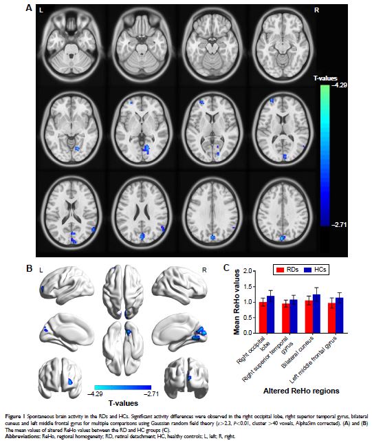
Results: Compared with HCs, RD patients had significantly decreased ReHo values
in the right occipital lobe, right superior temporal gyrus, bilateral cuneus
and left middle frontal gyrus. Moreover, we found that the mean ReHo signal of
the bilateral cuneus showed positive relationships with the duration of the RD
(r =0.392, P =0.032).
Conclusion: The RD patients showed brain neural homogeneity dysfunction in the
visual pathway, which may underline the pathological mechanism of RD patients
with acute vision loss. Besides, the ReHo values can reflect the progress of
the RD disease.
Keywords: retinal detachment, neural regional homogeneity, resting state,
functional magnetic resonance imaging