119370
论文已发表
注册即可获取德孚的最新动态
IF 收录期刊
- 3.6 Breast Cancer (Dove Med Press)
- 4.3 Clin Epidemiol
- 2.6 Cancer Manag Res
- 3.2 Infect Drug Resist
- 4.1 Clin Interv Aging
- 6.1 Drug Des Dev Ther
- 4.1 Int J Chronic Obstr
- 8.7 Int J Nanomed
- 2.5 Int J Women's Health
- 3.2 Neuropsych Dis Treat
- 2.4 OncoTargets Ther
- 2.6 Patient Prefer Adher
- 2.6 Ther Clin Risk Manag
- 3.1 J Pain Res
- 3.5 Diabet Metab Synd Ob
- 4.5 Psychol Res Behav Ma
- 3.4 Nat Sci Sleep
- 2.4 Pharmgenomics Pers Med
- 2.6 Risk Manag Healthc Policy
- 4.6 J Inflamm Res
- 2.3 Int J Gen Med
- 3.9 J Hepatocell Carcinoma
- 3.3 J Asthma Allergy
- 2.5 Clin Cosmet Investig Dermatol
- 3.0 J Multidiscip Healthc

HPIP: 淋巴结转移和子宫颈癌生存率差的预测因子
Authors Cao S, Sun J, Lin S, Zhao L, Wu D, Liang T, Sheng W
Received 6 May 2017
Accepted for publication 25 June 2017
Published 26 August 2017 Volume 2017:10 Pages 4205—4211
DOI https://doi.org/10.2147/OTT.S141248
Checked for plagiarism Yes
Review by Single-blind
Peer reviewers approved by Dr Akshita Wason
Peer reviewer comments 3
Editor who approved publication: Dr XuYu Yang
Background: The aim of this study was to explore the relationships of HPIP
expression status with the clinicopathological variables and survival outcomes
of patients with cervical cancer (CC).
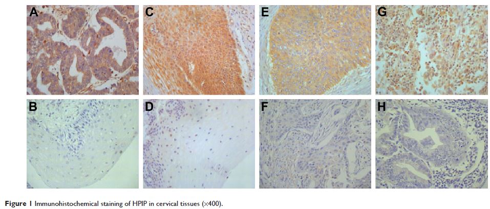
Methods: We compared the HPIP expression of 119 samples from CC tissues, 20 from
cervical intraepithelial tissues, and 20 from normal cervical tissues by using
immunohistochemical staining.
Results: It was observed that the ratio of elevated HPIP expression was
higher in CC tissues than in cervical intraepithelial neoplasia (P =0.017) and normal cervical
tissues (P =0.001). In addition, there was
an association between HPIP and clinicopathological factors, such as
histological grade (P <0.001),
stromal infiltration (P =0.015), lymph
node metastasis (P <0.001), lymphovascular space
invasion (LVSI; P =0.026), and
recurrence (P =0.029). Furthermore,
multivariate Cox regression analysis revealed that high HPIP expression (P =0.027 and P =0.042) as well as
the International Federation of Gynaecology and Obstetrics stage (P =0.003 and P =0.009), lymph node metastasis (P =0.031 and P =0.017), and LVSI (P =0.024 and P =0.046) were independent
prognostic factors. In addition, we demonstrated that high HPIP expression (P =0.003) and LVSI (P <0.001) were independently
related to lymph node metastasis.
Conclusion: Elevated HPIP expression may contribute to the progression and
metastasis of CC and may also serve as a new biomarker to predict the prognosis
of CC.
Keywords: HPIP, cervical cancer, metastasis, progression, prognosis