109451
论文已发表
注册即可获取德孚的最新动态
IF 收录期刊
- 3.4 Breast Cancer (Dove Med Press)
- 3.2 Clin Epidemiol
- 2.6 Cancer Manag Res
- 2.9 Infect Drug Resist
- 3.7 Clin Interv Aging
- 5.1 Drug Des Dev Ther
- 3.1 Int J Chronic Obstr
- 6.6 Int J Nanomed
- 2.6 Int J Women's Health
- 2.9 Neuropsych Dis Treat
- 2.8 OncoTargets Ther
- 2.0 Patient Prefer Adher
- 2.2 Ther Clin Risk Manag
- 2.5 J Pain Res
- 3.0 Diabet Metab Synd Ob
- 3.2 Psychol Res Behav Ma
- 3.4 Nat Sci Sleep
- 1.8 Pharmgenomics Pers Med
- 2.0 Risk Manag Healthc Policy
- 4.1 J Inflamm Res
- 2.0 Int J Gen Med
- 3.4 J Hepatocell Carcinoma
- 3.0 J Asthma Allergy
- 2.2 Clin Cosmet Investig Dermatol
- 2.4 J Multidiscip Healthc

扩增光谱参数与小鼠结肠直肠肿瘤同种移植的组织病理学指标相关联的初步体验
Authors Zhang XY, Li XT, Sun J, Sun YS
Received 10 November 2016
Accepted for publication 16 May 2017
Published 26 August 2017 Volume 2017:10 Pages 4213—4223
DOI https://doi.org/10.2147/OTT.S127283
Checked for plagiarism Yes
Review by Single-blind
Peer reviewers approved by Dr Amy Norman
Peer reviewer comments 3
Editor who approved publication: Dr Carlos Vigil Gonzales
Purpose: To determine the correlation between continuously distributed
diffusion-weighted image (DWI)-derived parameters and histopathologic indexes.
Methods: Fifty-four mice bearing HCT-116 colorectal
tumors were included for analysis; 12 mice were used for continuous
observation, and the other 42 mice were used for break-point observation. All
mice were randomly divided into radiotherapy and non-radiotherapy groups.
Optical imaging and MRI were performed at different time points according to
radiotherapy regimen (baseline, 24 h, 48 h, 72 h, 7 d, 14 d, and 28 d).
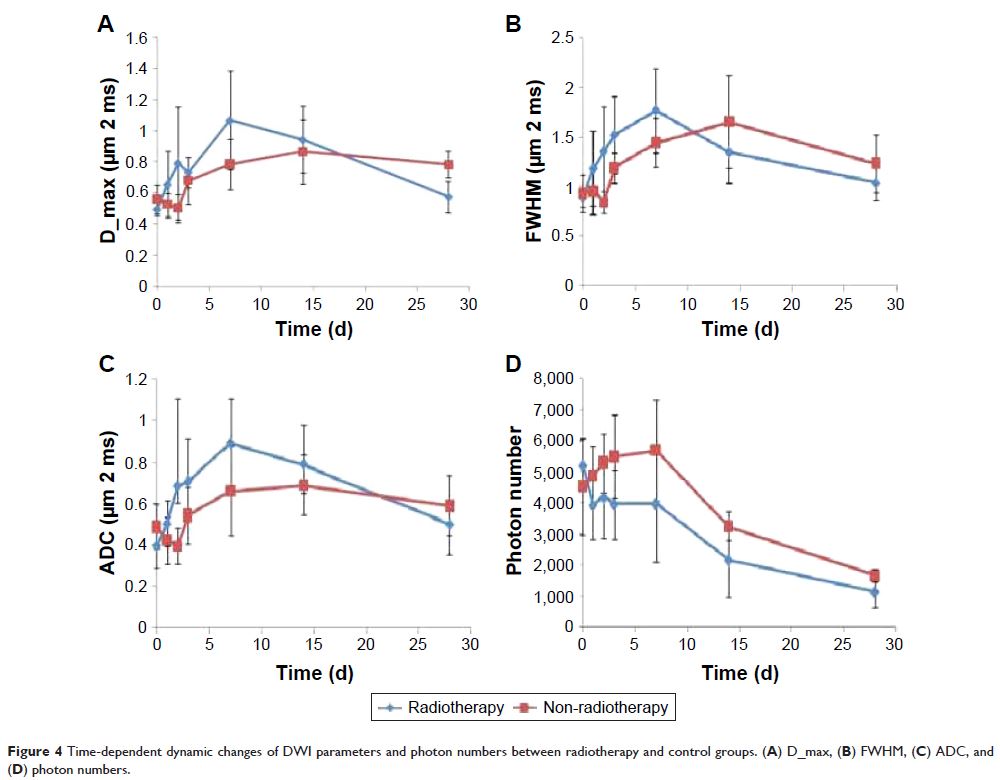
Continuous observation data were analyzed to show the difference of dynamic
changing trends of optical and MR-DWI–derived parameters between radiotherapy
and non-radiotherapy groups (photon numbers, D_max, full width half maximum
[FWHM], and apparent diffusion coefficient [ADC] value). Break-point observation
data were used to analyze the correlation between histopathologic indices and
DWI-derived parameters.
Results: There was a significant difference in the
changing trends of photon numbers, D_max, FWHM, and ADC value between
radiotherapy and non-radiotherapy groups, especially at early time points.
There was moderate negative correlation between Ki67 and percentage changes of
D_max, FWHM, and ADC values (the correlation coefficients were 0.632, 0.449,
and 0.586, P <0.001, P =0.008, and P <0.001, respectively). There
was moderate negative correlation between survivin and percentage changes of
D_max and ADC values (correlation coefficients were 0.496 and 0.473, P =0.004 and P =0.006, respectively).
Conclusion: The continuously distributed DWI-derived
parameters could reflect histological behavior to some extent and, thus, are
potential markers for early noninvasive monitoring of tumor cell apoptosis and
proliferation.
Keywords: magnetic
resonance imaging, diffusion-weighted imaging, continuously distributed, colorectal
cancer, murine homografts
