109451
论文已发表
注册即可获取德孚的最新动态
IF 收录期刊
- 3.4 Breast Cancer (Dove Med Press)
- 3.2 Clin Epidemiol
- 2.6 Cancer Manag Res
- 2.9 Infect Drug Resist
- 3.7 Clin Interv Aging
- 5.1 Drug Des Dev Ther
- 3.1 Int J Chronic Obstr
- 6.6 Int J Nanomed
- 2.6 Int J Women's Health
- 2.9 Neuropsych Dis Treat
- 2.8 OncoTargets Ther
- 2.0 Patient Prefer Adher
- 2.2 Ther Clin Risk Manag
- 2.5 J Pain Res
- 3.0 Diabet Metab Synd Ob
- 3.2 Psychol Res Behav Ma
- 3.4 Nat Sci Sleep
- 1.8 Pharmgenomics Pers Med
- 2.0 Risk Manag Healthc Policy
- 4.1 J Inflamm Res
- 2.0 Int J Gen Med
- 3.4 J Hepatocell Carcinoma
- 3.0 J Asthma Allergy
- 2.2 Clin Cosmet Investig Dermatol
- 2.4 J Multidiscip Healthc

负载紫杉醇 (Paclitaxel) 并以 A10-3.2 适体为靶向的聚 (丙交酯 - co - 乙醇酸) 纳米气泡用于超声成像和前列腺癌的治疗
Authors Wu M, Wang Y, Wang YR, Zhang MB, Luo YK, Tang J, Wang ZG, Wang D, Hao L, Wang ZB
Received 3 March 2017
Accepted for publication 30 May 2017
Published 26 July 2017 Volume 2017:12 Pages 5313—5330
DOI https://doi.org/10.2147/IJN.S136032
Checked for plagiarism Yes
Review by Single-blind
Peer reviewers approved by Dr Lakshmi Kiran Chelluri
Peer reviewer comments 3
Editor who approved publication: Dr Lei Yang
Abstract: In the current study, we synthesized prostate cancer-targeting
poly(lactide-co -glycolic acid) (PLGA)
nanobubbles (NBs) modified using A10-3.2 aptamers targeted to prostate-specific
membrane antigen (PSMA) and encapsulated paclitaxel (PTX). We also investigated
their impact on ultrasound (US) imaging and therapy of prostate cancer.
PTX-A10-3.2-PLGA NBs were developed using water-in-oil-in-water
(water/oil/water) double emulsion and carbodiimide chemistry approaches.
Fluorescence imaging together with flow cytometry verified that the
PTX-A10-3.2-PLGA NBs were successfully fabricated and could specifically bond
to PSMA-positive LNCaP cells. We speculated that, in vivo, the PTX-A10-3.2-PLGA
NBs would travel for a long time, efficiently aim at prostate cancer cells, and
sustainably release the loaded PTX due to the improved permeability together
with the retention impact and US-triggered drug delivery. The results demonstrated
that the combination of PTX-A10-3.2-PLGA NBs with low-frequency US achieved
high drug release, a low 50% inhibition concentration, and significant cell
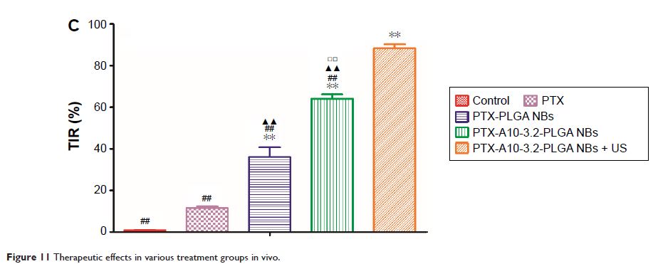
apoptosis in vitro. For mouse prostate tumor xenografts, the use of
PTX-A10-3.2-PLGA NBs along with low-frequency US achieved the highest tumor
inhibition rate, prolonging the survival of tumor-bearing nude mice without
obvious systemic toxicity. Moreover, LNCaP xenografts in mice were utilized to
observe modifications in the parameters of PTX-A10-3.2-PLGA and PTX-PLGA NBs in
the contrast mode and the allocation of fluorescence-labeled PTX-A10-3.2-PLGA
and PTX-PLGA NBs in live small animals and laser confocal scanning microscopy
fluorescence imaging. These results demonstrated that PTX-A10-3.2-PLGA NBs showed
high gray-scale intensity and aggregation ability and showed a notable signal
intensity in contrast mode as well as aggregation ability in fluorescence
imaging. In conclusion, we successfully developed an A10-3.2 aptamer and loaded
PTX-PLGA multifunctional theranostic agent for the purpose of obtaining US
images of prostate cancer and providing low-frequency US-triggered therapy of
prostate cancer that was likely to constitute a strategy for both prostate
cancer imaging and chemotherapy.
Keywords: nanobubbles, ultrasound imaging, paclitaxel, cancer therapy, aptamer,
prostate-specific membrane antigen
