109229
论文已发表
注册即可获取德孚的最新动态
IF 收录期刊
- 3.4 Breast Cancer (Dove Med Press)
- 3.2 Clin Epidemiol
- 2.6 Cancer Manag Res
- 2.9 Infect Drug Resist
- 3.7 Clin Interv Aging
- 5.1 Drug Des Dev Ther
- 3.1 Int J Chronic Obstr
- 6.6 Int J Nanomed
- 2.6 Int J Women's Health
- 2.9 Neuropsych Dis Treat
- 2.8 OncoTargets Ther
- 2.0 Patient Prefer Adher
- 2.2 Ther Clin Risk Manag
- 2.5 J Pain Res
- 3.0 Diabet Metab Synd Ob
- 3.2 Psychol Res Behav Ma
- 3.4 Nat Sci Sleep
- 1.8 Pharmgenomics Pers Med
- 2.0 Risk Manag Healthc Policy
- 4.1 J Inflamm Res
- 2.0 Int J Gen Med
- 3.4 J Hepatocell Carcinoma
- 3.0 J Asthma Allergy
- 2.2 Clin Cosmet Investig Dermatol
- 2.4 J Multidiscip Healthc

已发表论文
利用碘化油与包被荧光介孔二氧化硅的氧化铁纳米颗粒进行磁共振成像/计算机断层扫描/荧光三模造影
Authors Xue S, Wang Y, Wang M, Zhang L, Du X, Gu H, Zhang C
Published Date May 2014 Volume 2014:9(1) Pages 2527—2538
DOI http://dx.doi.org/10.2147/IJN.S59754
Received 26 December 2013, Accepted 15 March 2014, Published 21 May 2014
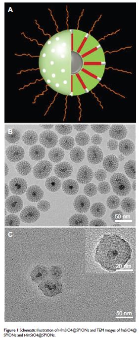
Abstract: In this study, a novel magnetic resonance imaging (MRI)/computed tomography (CT)/fluorescence trifunctional probe was prepared by loading iodinated oil into fluorescent mesoporous silica-coated superparamagnetic iron oxide nanoparticles (i-fmSiO4@SPIONs). Fluorescent mesoporous silica-coated superparamagnetic iron oxide nanoparticles (fmSiO4@SPIONs) were prepared by growing fluorescent dye-doped silica onto superparamagnetic iron oxide nanoparticles (SPIONs) directed by a cetyltrimethylammonium bromide template. As prepared, fmSiO4@SPIONs had a uniform size, a large surface area, and a large pore volume, which demonstrated high efficiency for iodinated oil loading. Iodinated oil loading did not change the sizes of fmSiO4@SPIONs, but they reduced the MRI T2 relaxivity (r2) markedly. I-fmSiO4@SPIONs were stable in their physical condition and did not demonstrate cytotoxic effects under the conditions investigated. In vitro studies indicated that the contrast enhancement of MRI and CT, and the fluorescence signal intensity of i-fmSiO4@SPION aqueous suspensions and macrophages, were intensified with increased i-fmSiO4@SPION concentrations in suspension and cell culture media. Moreover, for the in vivo study, the accumulation of i-fmSiO4@SPIONs in the liver could also be detected by MRI, CT, and fluorescence imaging. Our study demonstrated that i-fmSiO4@SPIONs had great potential for MRI/C/fluorescence trimodal imaging.
Keywords: multifunctional probe, SPIONs, mesoporous silica, iodinated oil
Keywords: multifunctional probe, SPIONs, mesoporous silica, iodinated oil
