109568
论文已发表
注册即可获取德孚的最新动态
IF 收录期刊
- 3.4 Breast Cancer (Dove Med Press)
- 3.2 Clin Epidemiol
- 2.6 Cancer Manag Res
- 2.9 Infect Drug Resist
- 3.7 Clin Interv Aging
- 5.1 Drug Des Dev Ther
- 3.1 Int J Chronic Obstr
- 6.6 Int J Nanomed
- 2.6 Int J Women's Health
- 2.9 Neuropsych Dis Treat
- 2.8 OncoTargets Ther
- 2.0 Patient Prefer Adher
- 2.2 Ther Clin Risk Manag
- 2.5 J Pain Res
- 3.0 Diabet Metab Synd Ob
- 3.2 Psychol Res Behav Ma
- 3.4 Nat Sci Sleep
- 1.8 Pharmgenomics Pers Med
- 2.0 Risk Manag Healthc Policy
- 4.1 J Inflamm Res
- 2.0 Int J Gen Med
- 3.4 J Hepatocell Carcinoma
- 3.0 J Asthma Allergy
- 2.2 Clin Cosmet Investig Dermatol
- 2.4 J Multidiscip Healthc

256 层螺旋 CT 扫描血管造影在对胃癌患者周围动脉进行术前评价中的优势
Authors Wu D, Zhao L, Liu Y, Wang J, Hu W, Feng X, Lv Z, Li Y, Yao X
Received 10 May 2015
Accepted for publication 30 June 2016
Published 16 February 2017 Volume 2017:10 Pages 927—933
DOI https://doi.org/10.2147/OTT.S88330
Checked for plagiarism Yes
Review by Single-blind
Peer reviewers approved by Dr Dekuang Zhao
Peer reviewer comments 3
Editor who approved publication: Dr Jianmin Xu
Objective: To evaluate the utilization of 256-slice spiral computed tomography (CT)
angiography in preoperative assessment of perigastric vascular anatomy in
patients with gastric cancer.
Methods: In this study, 80 gastric cancer patients were
included. The medical procedure of 256-slice spiral CT angiography was performed
on each of these patients consecutively. Thereafter, these patients were
subjected to surgical treatment in our hospital. The techniques of volume
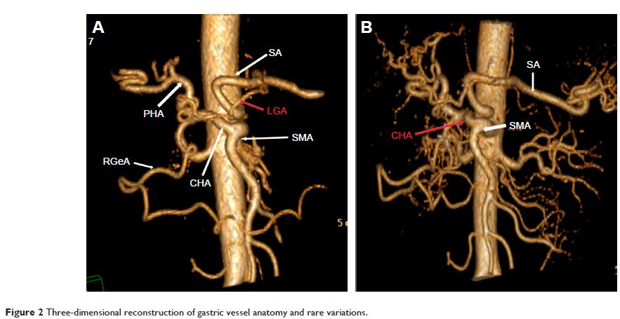
rendering (VR) and maximum intensity projection (MIP) were used to image
reconstruction of arteries around the stomach.
Results: Both VR and MIP were applied to reconstruct the images
of perigastric arteries. The results indicated that VR imaging was inferior to
MIP in determining the variant small artery anatomy around the greater
curvature and fundus. The respective rates of imaging produced by VR and MIP
for left gastroepiploic artery, short gastric artery, and posterior gastric
artery, were 32.50% versus 100%, 16.25% versus 87.50%, and 3.75% versus 25.00%,
respectively. According to Hiatt’s classification, 75 out of 240 cases were
abnormal types, among which we found Type II in 30 cases, Type III in 33 cases,
Type IV in three cases, Type V in six cases, and Type VI in only three cases.
There was no significant difference for total and every single variation type,
between our group and Hiatt’s group (P >0.05).
Conclusion: The 256-slice spiral CT angiography can be regarded as
an effective and accurate diagnostic modality for preoperative assessing
anatomical arterial variations in gastric cancer; MIP was superior to VR at
identifying variations of some small artery, whereas VR was better than MIP at
showing anatomical arterial variations due to its three-dimensional effect.
Keywords: gastric cancer, artery, angiography,
tomography, spiral computed
