109568
论文已发表
注册即可获取德孚的最新动态
IF 收录期刊
- 3.4 Breast Cancer (Dove Med Press)
- 3.2 Clin Epidemiol
- 2.6 Cancer Manag Res
- 2.9 Infect Drug Resist
- 3.7 Clin Interv Aging
- 5.1 Drug Des Dev Ther
- 3.1 Int J Chronic Obstr
- 6.6 Int J Nanomed
- 2.6 Int J Women's Health
- 2.9 Neuropsych Dis Treat
- 2.8 OncoTargets Ther
- 2.0 Patient Prefer Adher
- 2.2 Ther Clin Risk Manag
- 2.5 J Pain Res
- 3.0 Diabet Metab Synd Ob
- 3.2 Psychol Res Behav Ma
- 3.4 Nat Sci Sleep
- 1.8 Pharmgenomics Pers Med
- 2.0 Risk Manag Healthc Policy
- 4.1 J Inflamm Res
- 2.0 Int J Gen Med
- 3.4 J Hepatocell Carcinoma
- 3.0 J Asthma Allergy
- 2.2 Clin Cosmet Investig Dermatol
- 2.4 J Multidiscip Healthc

Axl 促进维尔姆斯肿瘤 (Wilms' tumor) 的增殖、侵袭和迁移,并可用作预后因子
Authors Zhu S, Liu G, Fu W, Hu J, Fu K, Jia W
Received 11 November 2016
Accepted for publication 5 January 2017
Published 16 February 2017 Volume 2017:10 Pages 955—963
DOI https://doi.org/10.2147/OTT.S127419
Checked for plagiarism Yes
Review by Single-blind
Peer reviewers approved by Dr Colin Mak
Peer reviewer comments 2
Editor who approved publication: Dr Ingrid Espinoza
Purpose: Overexpression of Axl has
been reported in many tumors, where it promotes tumorigenesis and progression,
as well as correlates with the prognosis of different malignancies. However, Axl expression and its function have
rarely been reported in Wilms’ tumor (WT). This study aimed to reveal the
clinical significance of Axl expression
in patients with WT and determine its mechanisms.
Materials and methods: We analyzed the expression of Axl and its
correlations with various clinicopathological features in 72 WT tissues and 72
adjacent non-cancerous tissues by immunohistochemistry. Cox proportional
hazards regression models were used to investigate the correlations between Axl expression
and the prognosis of WT patients. Fresh frozen samples from 20 WT patients were
examined using Western blotting (WB) and real-time quantitative polymerase
chain reaction (RT-qPCR). In WT cell line, after Axl knockdown
by sh-Axl and growth arrest-specific 6 (Gas6)
stimulation, the cell proliferation, migration and invasion abilities were
detected by methyl-thiazolyl-tetrazolium (MTT), clone-forming, wound-healing
and transwell assays. Meanwhile, the tumor-forming ability was tested on nude
mice xenograft models. Finally, the expression of several proteins in signal
pathways was quantified by WB assays.
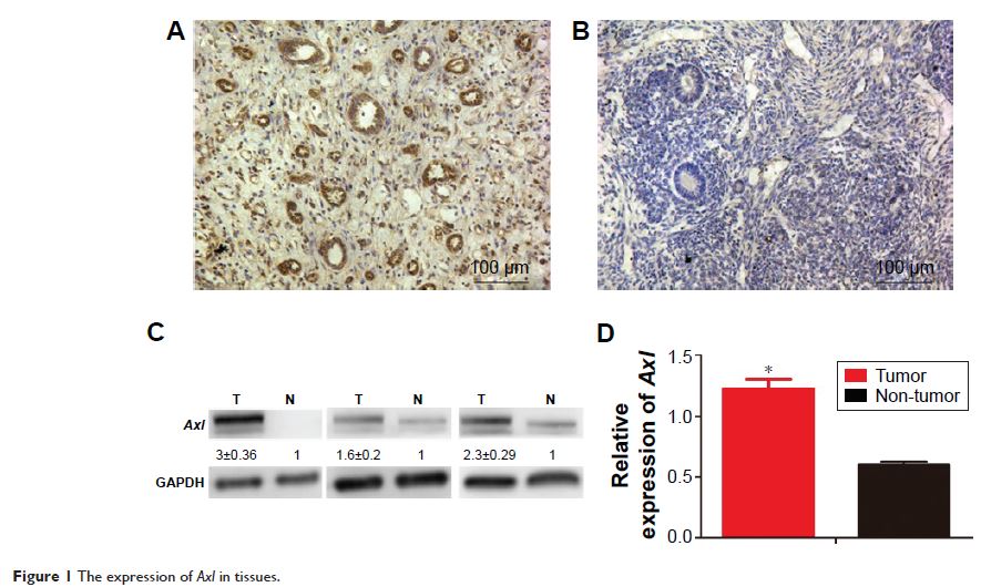
Results: Compared with the adjacent non-cancerous tissues, the
expression of Axl was
significantly higher in WT tissues (P <0.05). High
expression of Axl was
associated with tumor recurrence or lung metastasis of WT patients and was a prognostic
factor for WT patients (P <0.05). In
vitro assays, the proliferation, migration and invasion of WT cells decreased
with Axl knockdown and significantly increased
with Axl activation
by Gas6 (P <0.05). In vivo assays,
the ability of tumorigenicity in WT cells reduced dramatically after Axl knockout
(P <0.05). Moreover, PI3K–Akt pathway proteins decreased with Axl knockdown.
Conclusion: Our results suggest that Axl is
highly expressed in WT and is a prognostic factor, which could promote the
progression of WT in vitro and in vivo. It may also be a potential biomarker
for WT.
Keywords: Wilms’
tumor, Axl , prognosis, proliferation,
invasion, migration
