109814
论文已发表
注册即可获取德孚的最新动态
IF 收录期刊
- 3.4 Breast Cancer (Dove Med Press)
- 3.2 Clin Epidemiol
- 2.6 Cancer Manag Res
- 2.9 Infect Drug Resist
- 3.7 Clin Interv Aging
- 5.1 Drug Des Dev Ther
- 3.1 Int J Chronic Obstr
- 6.6 Int J Nanomed
- 2.6 Int J Women's Health
- 2.9 Neuropsych Dis Treat
- 2.8 OncoTargets Ther
- 2.0 Patient Prefer Adher
- 2.2 Ther Clin Risk Manag
- 2.5 J Pain Res
- 3.0 Diabet Metab Synd Ob
- 3.2 Psychol Res Behav Ma
- 3.4 Nat Sci Sleep
- 1.8 Pharmgenomics Pers Med
- 2.0 Risk Manag Healthc Policy
- 4.1 J Inflamm Res
- 2.0 Int J Gen Med
- 3.4 J Hepatocell Carcinoma
- 3.0 J Asthma Allergy
- 2.2 Clin Cosmet Investig Dermatol
- 2.4 J Multidiscip Healthc

Lumivascular(血管内成像)光学相干断层扫描引导下的粥样斑块切除术用于支架内再狭窄相关的复发性股腘动脉闭塞性病变:病例系列报告
Authors Chan YC, Cheung GC, Cheng SW
Received 11 May 2020
Accepted for publication 8 July 2020
Published 29 July 2020 Volume 2020:16 Pages 325—329
DOI https://doi.org/10.2147/VHRM.S260190
Checked for plagiarism Yes
Review by Single anonymous peer review
Peer reviewer comments 3
Editor who approved publication: Dr Harry Struijker-Boudier
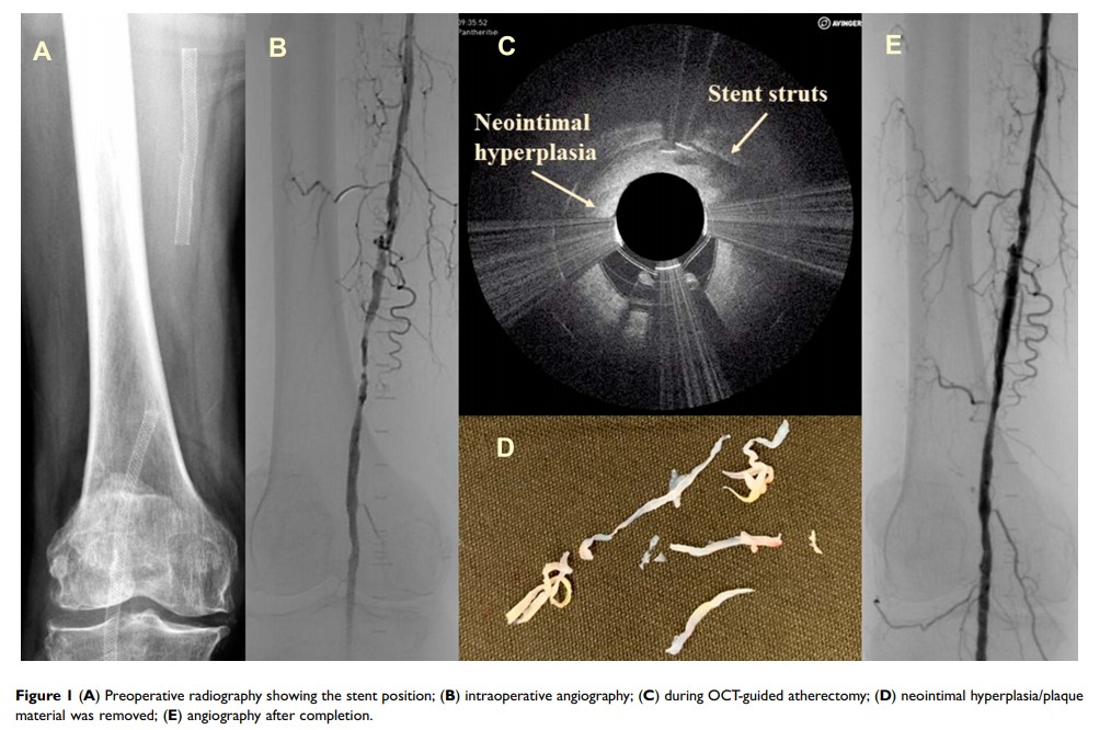
Abstract: Lumivascular optical coherence tomography (OCT) is a novel adjunct in the field of medicine. It offers clear real-time imaging of artery walls before and during endovascular intervention. This study reports our initial experience on the use of lumivascular OCT-guided atherectomy in the management of two patients with recurrent restenosis in their femoropopliteal arteries associated with in-stent restenosis. Endovascular procedures were successful with a Pantheris atherectomy device (Avinger, Redwood City, CA, USA) and drug-eluting balloons. The OCT images clearly distinguished normal anatomy from plaque pathology, were of great advantage in both the accurate diagnosis and treatment of target lesions, and may reduce radiation during the endovascular procedure. However, the price of the device and its need for contrast infusion limit its routine clinical use.
Keywords: optical coherence tomography, peripheral arterial disease, vascular, endovascular, diagnosis, intervention, atherectomy
